Mechanisms of “Fetomaternal Tolerance” and the Immunology of the Maternal-fetal Interface
The mystery of how the fetus avoids rejection by the maternal immune system has fascinated both reproductive biologists and immunologists alike for over 60 years. The fact that underlying mechanisms have remained obscure for so long is all the more remarkable given that pregnancy formally demonstrates the possibility of establishing immune tolerance to entire organs. We have identified several of the major mechanisms of fetomaternal tolerance in mice. These include restrictions on dendritic cell trafficking from the pregnant uterus (Collins et al., J. Clin. Invest., 2009), limitations on the number of dendritic cells and macrophages that are able to come near the placenta (Collins et al., J. Clin. Invest., 2009; Tagliani et al., J. Exp. Med. 2011), the shedding of non-immunogenic placental debris into maternal blood that fails to activate maternal T cells (Erlebacher et al., J. Clin. Invest., 2007; Tay et al., PLoS ONE, 2013), and the inability of the decidua, the specialized uterine stromal tissue that encases the conceptus, to recruited activated T cells from the blood and foster type 1 immune responses (Nancy et al., Science, 2012; Nancy et al., J. Clin. Invest., 2018). We are now studying these mechanisms in greater detail.

Mechanisms of fetomaternal tolerance – An Overview. This figure depicts much of what is currently appreciated about this phenomenon based upon the work of ours and others’ labs on maternal T cell responses to surrogate trophoblast antigens (e.g. transgenically expressed ovalbumin; Erlebacher et al., J. Clin. Invest., 2007). (1) Due to the low tissue density of dendritic cells and absence of lymphatic vessels within the decidua, maternal T cells become aware of trophoblast antigens exclusively as a result of their cell-free transport within the uterine lymphatic vessels and the maternal bloodstream, followed by their uptake and presentation by spleen- and lymph node-resident antigen-presenting cells. (2) Presentation of trophoblasts antigens is “non-immunogenic,” in that it induces an abortive proliferative response and fails to elicit effector T cell priming even when the mice are injected with adjuvants to induce costimulation. The causes of this response are currently incompletely understood, but in part involves the action of Treg cells. (3) Even if trophoblast antigen-specific T cells are activated via experimental means, the concept remains completely viable up through mid-gestation because the decidua excludes the cells from the maternal-fetal interface.

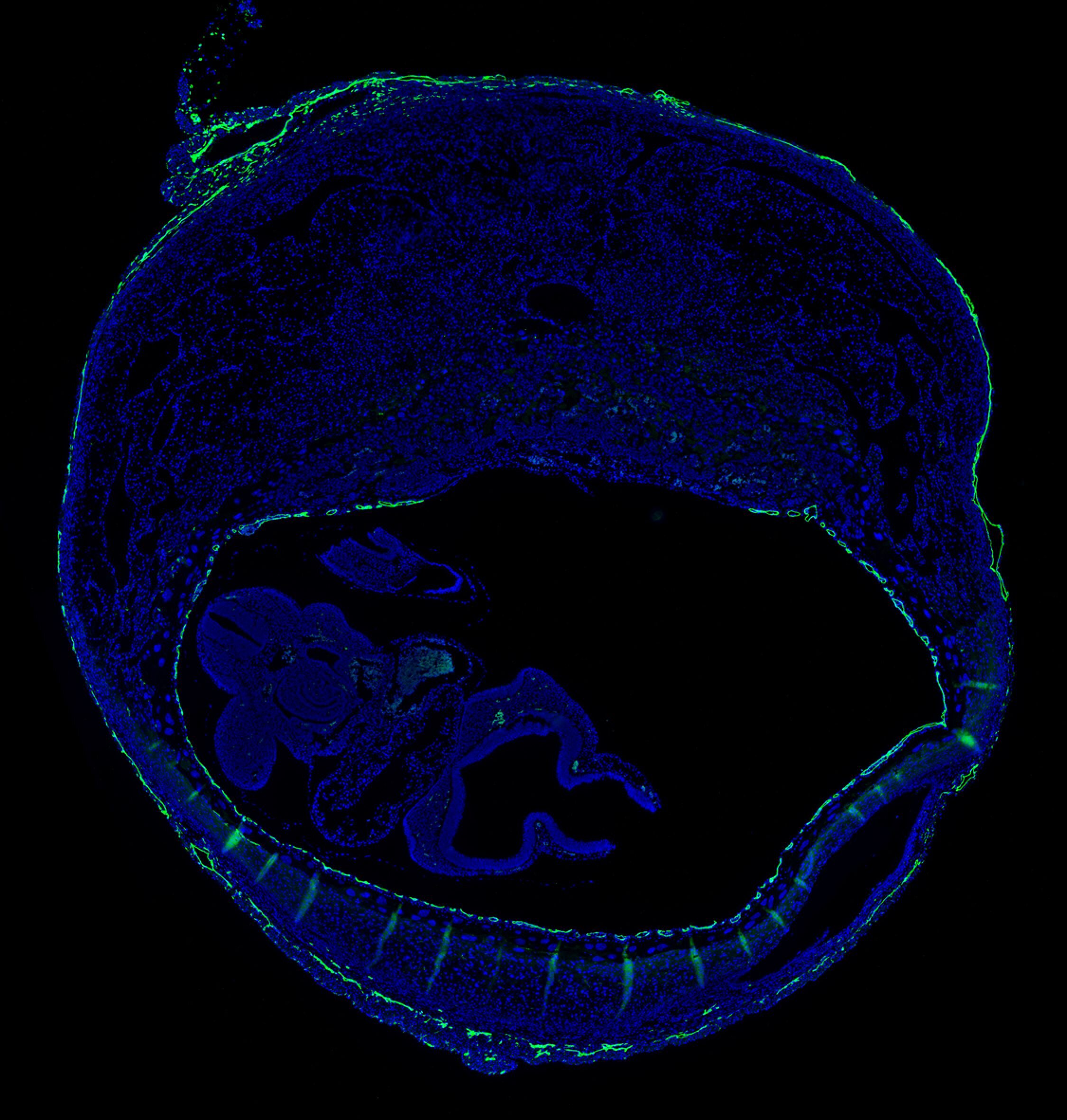
The decidua: a zone of immunological quiescence that excludes activated T cells. As a key mechanism for fetomaternal tolerance in mice that acts during the “effector phase” of the immune response, the decidua is unable to recruit activated Th1 cells and CTLs from the bloodstream. This can be visualized in mice that were pre-immunized to a protein antigen prior to pregnancy, and then systemically rechallenged with that same antigen soon after implantation. Decidual tissue sections were stained with an antibody to CD3 to visualize T cells (red). Note that the reactivated T cells accumulate in the myometrium but not the decidua. We have linked the exclusion of the T cells from the decidua to an inability of decidual stromal cells to express T cell-attracting chemokines, in turn, due to these genes’ epigenetic silencing via the repressive histone mark H3K27me3. From Nancy et al., Science (2012).